nerdyscience
New Members
-
Joined
-
Last visited
-
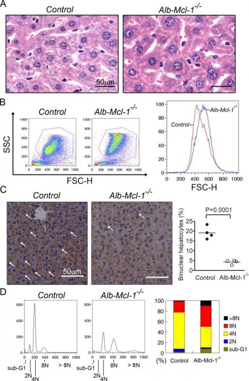
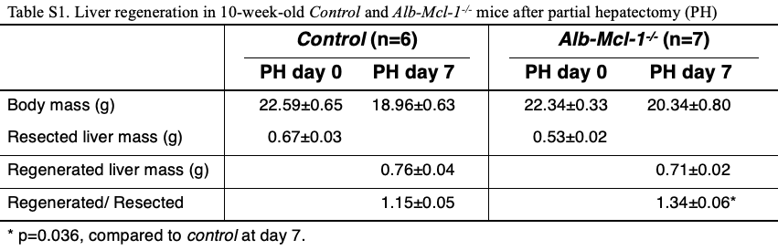
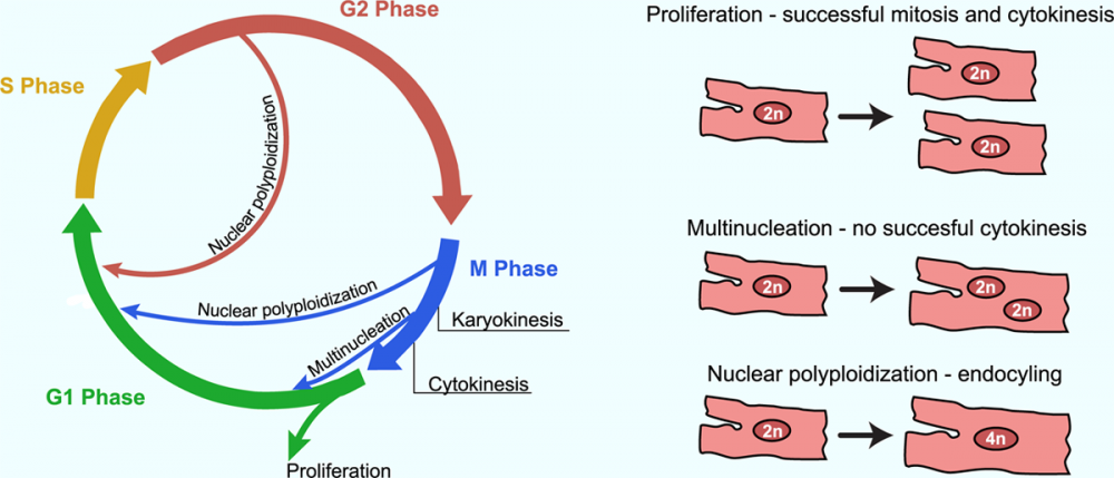
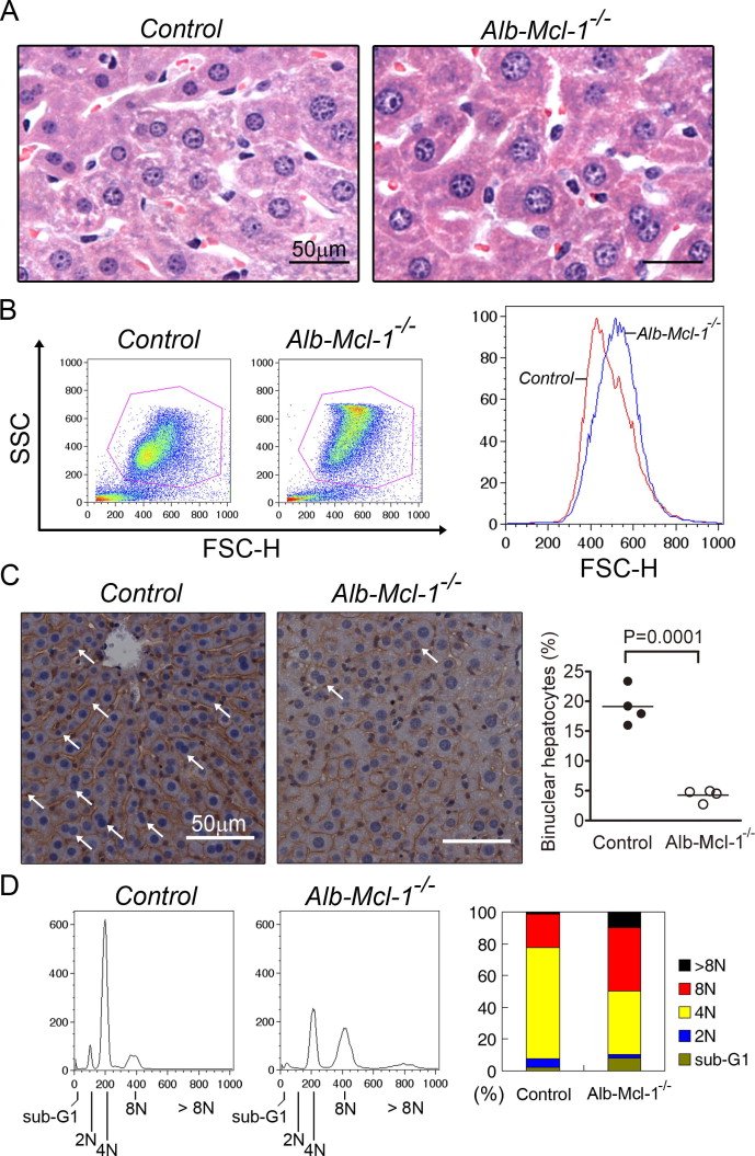
The emergence of polyploid cells in mammals can be associated with the development and differentiation of certain tissues. For instance, polyploid cells are present in the heart (cardiomyocytes: 4n), placenta (trophoblast giant cells: 8n to 64n), bone marrow (megakaryocytes: 16n to 128n), pancreas (acinar cells: 4n)19,20 and liver (hepatocytes: 4n to 8n)21,22,23. Ref: https://doi.org/10.1038/s41575-020-0284-x Figure 1. Multiple cell cycle variants are observed in mammalian cardiomyocytes. During normal productive cell cycle progression resulting in proliferation, the mother cell prepares itself during G1 phase and replicates its DNA during S phase. In G2 phase, the cell prepares for mitosis. During mitosis (M phase), the cell undergoes nuclear division (karyokinesis), and the 2 nuclei are separated in 2 daughter cells during cytokinesis. Alternatively, in cardiomyocytes, the cell cycle can also be abrogated after S phase but before karyokinesis (endocycling), resulting in a tetraploid nucleus. This process can repeat several times, leading to increased ploidy levels. Binucleated cells arise when the cell cycle abrogates after karyokinesis but before cytokinesis. Multiple successive rounds can also occur, leading to increased levels of multinucleation. The resulting cell types of these 2 variants are referred to as polyploid cells because they contain multiple copies of the original DNA content by having either multiple separate nuclei or one enlarged nucleus containing all the DNA. https://doi.org/10.1161/CIRCRESAHA.119.315408 Synergism between p53 and Mcl-1 in protecting from hepatic injury, fibrosis and cancer DOI: 10.1016/j.jhep.2010.07.035 Polyploid hepatocytes due to Mcl-1 knockout Partial hepatectomy (PH) analysis revealed that Mcl-1 knockout livers also regenerated better than control livers (Supplementary Table 1). Notably, Mcl-1 deficient hepatocytes tended to be enlarged and prone to undergo aberrant polyploidization (Supplementary Fig. 3). Supplementary Figure 3 Mcl-1 deficient hepatocytes manifest enlarged cell size and reduced binucleation. (A) Representative H & E staining of liver sections of 8-week-old control or Alb-Mcl-1-/- mice. (B) Primary hepatocytes isolated from 8-week-old control or Alb-Mcl-1-/- mice were stained with propidium iodide and analyzed by flow cytometry. The forward scatter height (FSC-H) distributions of the two gated hepatocyte populations as indicated in the left panel were plotted on the right panel. (C) Liver sections from 10-week-old control or Alb-Mcl-1-/- mice were stained with β-catenin antibody, and the percentage of binuclear hepatocyte (pointed by arrows) was calculated from at least 10 fields of 400X images for each mouse (n=4). (D) Flow cytometric analysis of the DNA content of primary hepatocytes from control or Alb-Mcl-1-/- mice at the age of 8-10 weeks. The result of one representative analysis is shown on the left panel. The average results from 4 pairs of mice are plotted on the right panel.